

ما الذي ستحصل عليه
52+
فيديوهات بالعربية
36+
مقالات طبية
300+
أسئلة حالات طبية
500+
بطاقات مراجعة
8 Tests
إمتحانات تقييمية لكل موضوع
1 Exam
إختبارات تقييمية
خطط دراسية جاهزة
تعلّم وتدرّب مع أفضل المعلمين الطبيين

خطط دراسية
خططنا الجاهزة للاستخدام تمنحك خارطة طريق واضحة تقسم المواد المعقدة إلى خطوات قابلة للإدارة، مما يبقيك مركزًا ومنظمًا وفي المسار الصحيح للنجاح في امتحانات USMLE وIMLE وNBME وغيرها.

محاضرات فيديو
تعلّم المفاهيم الطبية الأساسية من خلال دروس فيديو موجزة وجذابة، تُقدَّم باللغة العربية على يد مُعلمين طبيين ذوي خبرة. متاحة ضمن خطط الدراسة أو كمكتبة مستقلة.

صفحات ملخصة
وفّر وقتك بعد مشاهدة الفيديوهات من خلال ملخصات منظمة وسهلة المراجعة. غنية برسومات توضيحية عالية الجودة وفيديوهات وأسئلة تدريبية، جميعها مراجعة من قبل أطباء متخصصين.

بنك أسئلة وإمتحانات تقييمية
مارس ورسّخ تعلمك من خلال آلاف الحالات السريرية والتقييمات الذاتية الواقعية، مدعومة جميعها بشروحات مفصلة للإجابات ومحاضرات فيديو وصفحات ملخص موجزة.

بطاقات مراجعة
راجع بسرعة واحتفظ بالمعلومات عالية الأهمية باستخدام بطاقات تعليمية جاهزة للاستخدام، مصممة لمساعدتك على التركيز على ما يهم أكثر.
- ModuleReview ArticlesVideo LecturesPractice QuestionsFlashcards
ما يقوله المستخدمون
Lung Volumes and Capacities
Writer: Mustafa A. Shahrori, MD.
Keywords: Pulmonary function, spirometry, lung volumes, lung capacities, respiratory physiology, tidal volume, inspiratory reserve volume, expiratory reserve volume, residual volume, functional residual capacity, vital capacity, total lung capacity, pulmonary function testing, respiratory assessment
Overview
Lung volumes and capacities are fundamental respiratory measurements essential for diagnosing and monitoring pulmonary function. The four primary lung volumes (tidal volume, inspiratory reserve volume, expiratory reserve volume, and residual volume) represent discrete air quantities during different breathing phases. The four capacities (inspiratory capacity, functional residual capacity, vital capacity, and total lung capacity) combine these volumes. Measurement techniques, normal values, clinical patterns in obstructive and restrictive diseases, and physiological factors affecting these parameters provide crucial insights for respiratory assessment and diagnosis.
Definition and Classification
Pulmonary function tests (Figure 1) (Figure 2) (Figure 3) measure two primary categories of respiratory parameters:
1. Lung volumes - discrete, non-overlapping quantities of air in the lungs during different phases of the respiratory cycle
2. Lung capacities - combinations of two or more lung volumes

Figure 1. Pulmonary function test (PFT) procedure using spirometry

Figure 2. Lung volumes and capacities
Kapwatt. (2014, May 31). Added the “Inspiration Capacity” (IC) metric, which is the sum of Inspiratory Reserve Volume and Tidal Volume [Diagram]. Wikimedia Commons. https://commons.wikimedia.org/wiki/File:Lungvolumes_updated.svg
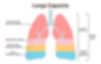
Figure 3. Lung volumes and capacities
The Four Lung Volumes
Tidal volume (VT)
The volume of air moved into or out of the lungs during normal quiet breathing
Typical value: 6-8 mL/kg of ideal body weight (approximately 500 mL in an average adult)
Measured during regular breathing without additional effort
NOTE: Tidal volume increases during exercise and can decrease during shallow breathing patterns such as those seen in restrictive lung diseases.
Inspiratory reserve volume (IRV)
The additional volume of air that can be inhaled with maximum effort beyond normal tidal inspiration
Typical value: 2.5-3.0 L in adults
Represents the reserve capacity for increased ventilation during exercise or respiratory distress
Expiratory reserve volume (ERV)
The additional volume of air that can be forcefully exhaled after a normal tidal expiration
Typical value: 1.0-1.5 L in adults
Decreased in obesity, pregnancy, and conditions causing abdominal distension
Residual volume (RV)
The volume of air remaining in the lungs after maximal expiration
Typical value: 1.0-1.5 L in adults
Cannot be voluntarily exhaled and requires indirect measurement techniques
Prevents complete lung collapse and maintains gas exchange between breaths
ALERT: Residual volume and any lung capacity that includes RV cannot be measured directly by simple spirometry. They require indirect measurement techniques such as nitrogen washout, helium dilution, or body plethysmography.
The Four Lung Capacities
Inspiratory capacity (IC)
Definition: IRV + VT
The maximum volume of air that can be inhaled after a normal quiet expiration
Typical value: 3.0-3.5 L in adults
Clinical significance: Decreased in restrictive lung diseases
Functional residual capacity (FRC)
Definition: ERV + RV
The volume of air present in the lungs at the end of normal quiet expiration
Typical value: 2.0-2.5 L in adults
Represents the point where the outward elastic recoil of the chest wall is balanced by the inward elastic recoil of the lungs
Vital capacity (VC)
Definition: IRV + VT + ERV
The maximum volume of air that can be exhaled after a maximum inspiration
Typical value: 4.0-5.0 L in adults, but varies with age, sex, height, and ethnicity
Often measured as forced vital capacity (FVC) during spirometry
Total lung capacity (TLC)
Definition: IRV + VT + ERV + RV, or simply VC + RV
The total volume of air in the lungs after maximal inspiration
Typical value: 5.5-6.0 L in adults
Clinical significance: Decreased in restrictive diseases, increased in obstructive diseases
Measurement Techniques
Spirometry
Measures directly: VT, IRV, ERV, VC, and IC
Cannot measure: RV, FRC, and TLC (since these include RV)
Most commonly used pulmonary function test
Results are typically expressed as absolute values and as percentages of predicted values based on age, sex, height, and ethnicity
NOTE: Spirometry (Figure 4) is the primary tool for diagnosing obstructive and restrictive lung diseases, monitoring disease progression, and evaluating response to therapy.

Figure 4. Spirometry
Jmarchn. (2013, June 10). Doing a spirometry [Photograph]. Wikimedia Commons. https://commons.wikimedia.org/wiki/File:DoingSpirometry.JPG
For a more detailed discussion, refer to the Spirometry article.
Gas dilution techniques
Used to measure FRC, which then allows calculation of RV and TLC
Methods include:
Nitrogen washout technique
Helium dilution method
Based on the principle of dilution of a known concentration of inert gas in a closed system
Body plethysmography
Uses Boyle's law (pressure × volume = constant) to determine all lung volumes and capacities
Most accurate method, especially in patients with air trapping or bullous disease
Measures the total volume of gas in the thoracic cavity, including trapped gas
Clinical Significance and Patterns of Abnormality
Obstructive lung diseases
Examples: Asthma, COPD, emphysema, chronic bronchitis
Pattern of abnormalities:
Increased RV and FRC due to air trapping
Normal or increased TLC
Decreased VC due to increased RV
Decreased forced expiratory flow rates
ALERT: In obstructive lung diseases, RV can increase by more than 300% of predicted values, leading to hyperinflation and increased work of breathing.
Restrictive lung diseases
Examples: Pulmonary fibrosis, pneumonia, acute respiratory distress syndrome, neuromuscular disorders
Pattern of abnormalities:
Decreased TLC
Proportionally decreased all lung volumes
Normal or increased FEV1/FVC ratio
Mixed pattern
Features of both obstructive and restrictive patterns
Examples: Bronchiectasis with fibrosis, cystic fibrosis
Factors Affecting Lung Volumes and Capacities
Physiological factors
1. Age
o TLC remains relatively stable until older age
o RV increases with age as elastic recoil decreases
o VC decreases with age
2. Sex
o Males typically have larger lung volumes than females of the same age and height
o Differences become apparent after puberty
3. Body size and position
o Taller individuals have larger lung volumes
o FRC decreases when changing from standing to supine position
o Obesity decreases ERV and FRC due to diaphragmatic displacement
NOTE: Lung volumes should always be interpreted with reference to predicted values that account for the individual's age, sex, height, and ethnicity.
Pathological conditions
1. Respiratory muscle weakness
o Decreased TLC, VC, and IC
o Normal or increased RV
2. Pregnancy
o Progressive decrease in FRC as pregnancy advances
o Relatively preserved VC and TLC
o Changes due to elevation of diaphragm by enlarging uterus
3. Altitude
o Increased ventilation at high altitude
o Increased TLC and VC with long-term adaptation
References
Guyton, A. C., & Hall, J. E. (2021). Guyton and Hall textbook of medical physiology (14th ed.). Elsevier.
عينات من محتوى الدورة
تتضمن جميع الباقات إمكانية الوصول الكامل إلى خطط الدراسة، ومحاضرات الفيديو، وصفحات الملخصات، وبنك الأسئلة، وبطاقات الذاكرة، ونظام حجز المدرسين الخصوصيين.
Free Offer
$مجانًا
1 Month
Text text text text 1
Text text text text 2
Text text text text 3
Text text text text 4
Text text text text 5
Self-Paced Program
$109.9
12 Months
47 درسًا مسجلاً
30 ورقة نقدية عالية العائد
أكثر من 300 سؤال
500 بطاقة تعليمية
2 التقييمات
1. اختبار تجريبي كامل
لوحة تحليلات البيانات.
Guided Program
$780.9
12 Months
دورة كاملة ذاتية التعلم
12 ساعة من الدروس الخصوصية
اجتماعان للتوجيه والإرشاد
خطة دراسية شخصية
قناة التواصل الخاصة بالمدرس
1. اختبار تجريبي كامل
دعم ذو أولوية


