
What You’ll Get
60+
Arabic Videos
73+
Concept Article
297+
Question Cases
718+
Smart Flashcards
1 Test
Module-based assessments
1 Exam
Evaluation exams
Pre-Built Study Plans
Learn and Practice With Top Medical Educators

Study Plans
Our ready-to-use plans give you a clear roadmap that breaks complex subjects into manageable steps, keeping you focused, organized, and on track to succeed in USMLE, IMLE, NBME, and more.

Video Lectures
Learn key medical concepts with concise, engaging video lessons, taught in Arabic by experienced medical educators. available with your study plans or as a standalone library.

Summary Pages
Save time after watching videos with organized and easy-to-review summaries. Packed with high-quality illustrations, videos, and practice questions, all reviewed by trained doctors.

QBank & Assessments
Practice and reinforce your learning with thousands of clinical cases and realistic self-assessments, all supported by in-depth answer explanations, video lectures, and concise summary pages.

Flashcards
Quickly review and retain high-yield information with ready-to-use flashcards, designed to help you focus on what matters most.
- ModuleReview ArticlesVideo LecturesPractice QuestionsFlashcardsAntimicrobials11945110Parasitology973389Virology141258137Mycology451739Bacteriology3527143343
What Users Say
The course was excellent. The doctors gave from their hearts and their goal was clear: to make every student understand and to provide every student with the information. They gave their maximum and did not fall short in anything.
Fadi Mari
The course was very, very good, beyond imagination. All the ideas presented at the beginning were well organized and helped a lot in understanding the material, solving questions, and progressing step by step. 🙏 All the doctors, without exception, presented amazing work, and their methods were easy to understand.
Islam Breek
Frankly, there are no words to describe the doctor. I thank him very much for his education and for teaching everything, big and small, explaining it from the bottom of his heart. Thank you very, very much for all the effort and care he gives every student.
Mohammad Yassin
I have to say that the effort, time, energy, and concern of Dr. Alaa for us to get the best out of the classes is just phenomenal. He makes sure every student understands the material, gives his maximum in teaching, and truly cares about our success in every way.
Rani Abu Arisheh
As for the doctors, they’re really excellent, their explanations are clear, and they make things super easy to understand. So far, every doctor I’ve had a course with was great and truly gave their all ❤️ The course itself is very good and professional, and the team is always ready to help 🙌
Mona Assadi
The dr’s are great—they explain everything straight to the point, making everything crystal clear, and they don’t move on until everyone is on track. The course itself is great, with high-yield material simplified for understanding. I already recommend it because it helps stay on track and makes learning easier.
Mustafa Sharara
A big and ambitious program with a lot of effort behind it. It’s led by top-level doctors who are almost always available to support us, and if anything is unclear, they help right away. Plus, amazing coordinators like Yasmin make the experience even better 🌟.
Abed Elwahab Abada
Honestly, the course is well-organized, from the materials to the schedule and both general and personal follow-up. Any small issue or question gets resolved within minutes 👍🏼👍🏼. The team is always available and supportive, making sure every student stays on track and gets the help they need to succeed.
Ahmad Judy
In short, a fully comprehensive program led by a highly experienced team. The program is very practical, strong, and effective, providing everything needed to succeed. The team is supportive and always available to help, making the learning experience smooth and ensuring every student gets the guidance they need.
Khaled Habib Alla
The program is awesome, I’m really happy 😍. The doctors are great, and the way they explain things is super clear 👌. Everything is well-organized, and the team is always ready to help. It makes learning easy, effective, and enjoyable, and I’m very grateful for all the support provided.
Naji Armaly
An excellent program with an outstanding teaching team 👏. The doctors are always available, explain everything clearly, and make learning enjoyable. The program is well-organized and provides all the support needed to succeed. I’m very grateful for the effort they put into helping every student reach their best.
Hmam Taha
Brocali means a lot to me. They are always available for us and help the students like brothers, which makes learning better and more exciting. I hope that one day I can make them proud of me, just as I am proud of everything they do for us.
Ajwad Mallak
The course is top! The doctors are amazing—huge thanks to them! Their explanations are clear, and the materials cover everything needed. I definitely recommend it strongly 💪. The best part is the team and management—they’re super helpful and always respond right away if anyone needs anything 🙌.
Orwa Abu Zeid
A big thank you to Brocali’s course for the personal support all the way until exam day, and for the clear plans made for each person to succeed in the exam. 🙏🏻 I really appreciate all your efforts and the care you put into helping every student succeed. 🙏🏻
Fawaz Mahajneh
Brocali is a very excellent platform. The doctors’ explanations are complete and detailed, making everything very useful and easy to understand. All due respect, may God bless you for your effort and dedication. The program truly helps every student succeed and provides all the support needed throughout the course.
Aya Khater
Brocali is the best medical educational institution. They simplify the most complicated material, making it easy to understand. I advise anyone to just sign up at Brocali, as I have tried many other medical teaching options and cannot compare them—they truly stand out and make learning effective and clear.
Khaled Asir
For me, Brocali helped me a lot in understanding all the concepts in very simple ways, thanks to the doctors knowing what is high-yield and what is not. Their explanations made studying much easier, and the course truly focuses on what we need to succeed in the exams.
Majd Taon
Brocali isn’t the only one, but it’s the best! I was guided by Brocali’s doctors and learned how to start my new medical journey, which changed many of my mindsets. I trusted Brocali as the way to succeed, and I’m very grateful for the time that introduced me to it.
Ameer Masarwa
Brocali provided me with all the knowledge and tools to pass the medical exams. They supported me through every step of the process, making sure I understood the material and stayed on track. Their guidance and dedication made preparing for the exams much easier and more effective.
Angham Mattar
Brocali played a major role in my life. They didn’t just prepare me for the exam; they also gave me the tools to navigate life’s obstacles. They cheered me up when things were hard and helped me become more confident in pursuing my passions and dreams.
Aseel Abu Al-Heja
I am a fifth-year medical student at DSMA, Ukraine. Preparing for Step 1, Brocali’s resources have been extremely helpful. I truly appreciate all the hard work this incredible team puts into Brocali. Thank you for making studying easier and providing the support every student needs to succeed.
Lina Hamza
Bacterial Stains and Cultures
Author: Adnan Mousa, M.D.
Keywords: Bacterial staining, Gram stain, Ziehl-Neelsen stain, special culture media, microbiology
Overview
Bacterial staining techniques are essential for identifying and differentiating microorganisms based on their structural and chemical properties. The Gram stain, the most widely used method, classifies bacteria as Gram-positive or Gram-negative based on their cell wall composition. Additional staining techniques address bacteria that do not stain effectively with Gram stain, such as acid-fast stains for Mycobacteria and Giemsa stain for intracellular organisms like Chlamydia and Plasmodium. Bacteria are further classified through biochemical tests, including catalase, coagulase, and hemolysis assays, aiding in distinguishing pathogenic species. Specialized stains like Periodic Acid–Schiff (PAS) highlight carbohydrate-rich structures, useful for diagnosing conditions such as Whipple disease.
Bacterial Staining Techniques
Bacterial staining is essential for differentiating microorganisms based on structural and chemical properties. The most commonly used stains in microbiology include:
Gram Stain
Gram staining is the first-line laboratory test for bacterial classification. It differentiates bacteria based on their cell wall structure.
Steps:
● Primary stain: First, a purple dye called crystal violet is applied to all the cells.
● Mordant: Next, iodine is added to help keep the purple dye in the cells.
● Decolorization: Then, alcohol or acetone is used to wash out the purple dye from Gram-negative bacteria.
● Counterstain: Finally, a pink dye called safranin is added, which stains the Gram-negative bacteria pink.
Interpretation:
● Gram-positive bacteria: Retain crystal violet stain due to a thick peptidoglycan layer and appear purple.
● Gram-negative bacteria: Lose the crystal violet stain and take up the counterstain (safranin), appearing red/pink.

Figure 1. Staph Aureus: A gram positive bacteria.
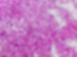
Figure 2. E. Coli: A gram negative bacteria.
Some bacteria do not stain effectively with Gram stain due to structural differences:
● Too thin to visualize: Treponema, Leptospira
● Lack a cell wall: Mycoplasma, Ureaplasma
● High lipid content in the cell wall: Mycobacteria
● Primarily intracellular or lack peptidoglycan: Legionella, Rickettsia, Chlamydia, Bartonella, Anaplasma, Ehrlichia
Gram-Positive Lab Algorithm
Gram-positive bacteria are classified based on their shape (bacilli or cocci), staining properties, and biochemical tests.
Gram-positive bacilli (rod-shaped, purple/blue staining):
Aerobic: These bacteria require oxygen to grow.
Listeria: Motile at room temperature, causes meningitis in newborns and pregnant women.
Bacillus: Forms spores; B. anthracis causes anthrax, B. cereus causes food poisoning.
Corynebacterium: Club-shaped; C. diphtheriae causes diphtheria (forms pseudomembrane in throat).
Anaerobic: These bacteria grow without oxygen.
Clostridium: Forms spores; C. tetani (tetanus), C. botulinum (botulism), C. perfringens (gas gangrene), C. difficile (pseudomembranous colitis).
Cutibacterium (formerly Propionibacterium): Part of normal skin flora; can cause acne and prosthetic joint infections.
Gram-positive cocci (circular, purple/blue staining):
● Branching filaments:
Nocardia: Weakly acid-fast (stains with acid-fast stain), causes pulmonary and cutaneous infections in immunocompromised patients.
Actinomyces: Not acid-fast, forms sulfur granules, causes abscesses (e.g., cervicofacial actinomycosis).
● Hemolysis on blood agar:
Alpha hemolysis (partial, green) (Figure 9):
- Streptococcus pneumoniae: Optochin-sensitive, bile-soluble, causes pneumonia and meningitis.
- Viridans streptococci: No capsule, part of normal oral flora, causes endocarditis.
Beta hemolysis (complete, clear):
- Group A (Streptococcus pyogenes): Bacitracin-sensitive, PYR-positive, causes pharyngitis, rheumatic fever, and glomerulonephritis.
- Group B (Streptococcus agalactiae): Bacitracin-resistant, causes neonatal meningitis and sepsis.
Gamma hemolysis (no hemolysis):
- Enterococcus: Grows in 6.5% NaCl, PYR-positive, causes urinary tract infections (UTIs) and endocarditis.
- Non-enterococcus (Streptococcus gallolyticus): Grows in bile, PYR-negative, associated with colon cancer and endocarditis.
Catalase test:
Catalase-positive (bubbles when hydrogen peroxide is added):
Staphylococcus: Forms clusters.
- S. aureus: Coagulase-positive, Novobiocin-resistant, causes skin infections, toxic shock syndrome, and food poisoning.
- S. epidermidis: Coagulase-negative, Novobiocin-sensitive, causes infections of prosthetic devices.
- S. saprophyticus: Novobiocin-resistant, causes UTIs in young women.
Catalase-begative (no bubbles):
- Streptococcus: Forms pairs or chains, further classified using PYR, Optochin, bile solubility, and Bacitracin tests.
Key tests explained:
● PYR test: Detects the enzyme pyrrolidonyl arylamidase.
○ Positive: Streptococcus pyogenes and Enterococcus.
○ Negative: Other streptococci.
● Optochin sensitivity: Differentiates S. pneumoniae (sensitive) from other alpha-hemolytic streptococci.
● Bile solubility: S. pneumoniae dissolves in bile; other streptococci do not.
● Bacitracin sensitivity: Differentiates Group A (S. pyogenes, sensitive) from Group B (S. agalactiae, resistant).
● Coagulase test: Differentiates S. aureus (positive) from other staphylococci (negative).
● Novobiocin sensitivity: Differentiates S. epidermidis (sensitive) from S. saprophyticus (resistant).
Gram-Negative Lab Algorithm
Key shapes:
● Diplococci (paired round cells):
○ Neisseria gonorrhoeae: Causes gonorrhea.
○ Neisseria meningitidis: Causes meningitis.
● Coccobacilli (short, oval-shaped rods):
○ Haemophilus influenzae: Causes respiratory infections and meningitis.
○ Bordetella pertussis: Causes whooping cough.
○ Pasteurella: Associated with animal bites.
○ Brucella: Causes brucellosis.
○ Francisella tularensis: Causes tularemia.
● Curved rods:
○ Campylobacter jejuni: Causes gastroenteritis.
○ Vibrio cholerae: Causes cholera.
○ Helicobacter pylori: Causes stomach ulcers and gastritis.
● Bacilli (rods):
○ Escherichia coli: Common cause of UTIs and diarrhea.
○ Klebsiella, Enterobacter, Citrobacter, Serratia: Opportunistic pathogens causing various infections.
○ Pseudomonas: Causes infections in immunocompromised patients.
○ Burkholderia: Includes pathogens like B. cepacia and B. pseudomallei.
○ Shigella: Causes dysentery.
○ Yersinia: Includes Y. pestis (plague) and Y. enterocolitica (gastroenteritis).
○ Salmonella: Causes typhoid fever and food poisoning.
○ Proteus: Known for urinary tract infections and swarming motility.
Growth characteristics:
● Aerobic: Grows in the presence of oxygen.
● Maltose fermentation: Seen in Neisseria meningitidis.
● Oxidase positive: Indicates the presence of cytochrome c oxidase enzyme.
○ Seen in Campylobacter jejuni, Vibrio cholerae, and Helicobacter pylori.
● Grows at 42°C: Campylobacter jejuni can tolerate higher temperatures.
● Grows in alkaline media: Vibrio cholerae thrives in alkaline conditions.
Fermentation characteristics:
● Lactose fermenters (fast-growing):
○ Escherichia coli, Klebsiella, Enterobacter: These bacteria ferment lactose quickly,
producing acid and gas.
● Lactose fermenters (slow-growing):
○ Citrobacter, Serratia: Ferment lactose more slowly.
● H2S production on TSI agar:
○ Salmonella, Proteus: Produce hydrogen sulfide (H2S), which turns the medium black.
Important tests:
● Oxidase test:
○ Positive in Neisseria gonorrhoeae, Moraxella, and Campylobacter jejuni.
○ Helps differentiate between oxidase-positive and oxidase-negative organisms.
● Urease production:
○ Seen in Helicobacter pylori, which breaks down urea to produce ammonia.
Important pathogens:
● Haemophilus influenzae: Causes respiratory and meningeal infections.
● Bordetella pertussis: Causes whooping cough.
● Neisseria gonorrhoeae: Causes gonorrhea.
● Pseudomonas: Opportunistic pathogen causing severe infections.
● Salmonella: Causes typhoid fever and gastroenteritis.
Giemsa Stain
Giemsa stain binds to nucleic acids, highlighting the nucleus and cytoplasm, making it ideal for visualizing intracellular organisms, certain extracellular bacteria, and protozoa. It produces purple-blue organisms against a lighter background, enhancing contrast for microscopic examination.
Common uses include detecting:
● Chlamydia spp. – Obligate intracellular bacteria lacking peptidoglycan, making Gram staining ineffective.
● Rickettsia spp. – Intracellular bacteria transmitted by arthropods (ticks, fleas, lice).
● Borrelia spp. – A large spirochete visible with Giemsa stain, responsible for Lyme disease and relapsing fever.
● Trypanosomes – Extracellular protozoa causing Chagas disease and African sleeping sickness, recognized by their flagellated morphology in blood smears.
● Helicobacter pylori: Spiral-shaped Gram-negative bacteria detected in gastric biopsies.
● Plasmodium spp. – Malaria parasites in RBCs, identified at various life stages (trophozoites, schizonts, gametocytes) in blood smears.
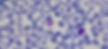
Figure 3. Gametocyte of plasmodium vivax on peripheral blood smear stained with giemsa.
Periodic Acid–Schiff (PAS) Stain
PAS stain detects glycogen, mucopolysaccharides, and other carbohydrate-rich structures, producing a magenta coloration. It is commonly used to highlight glycoproteins in basement membranes and fungal cell walls, and mucins in epithelial cells.
Clinical applications:
● Whipple disease (Tropheryma whipplei) – PAS-positive foamy macrophages in the lamina propria of the small intestine aid in diagnosis.
● Fungal infections – Stains the polysaccharide-rich cell walls of fungi, aiding in their identification.
● Glycogen storage diseases – Highlights intracellular glycogen deposits in affected tissues.

Figure 4. Whipple’s disease: intensely PAS-positive macrophages occupying the lamina propria mucosae.
Review Article Whipple's Disease: Our Own Experience and Review of the Literature - Scientific Figure on ResearchGate. Available from: https://www.researchgate.net/figure/Whipples-disease-intensely-PAS-positive-macrophages-occupying-the-lamina-propria_fig14_241060805 [accessed 11 Mar 2025]
Ziehl-Neelsen Stain (Carbol Fuchsin Stain)
The Ziehl-Neelsen (Carbol Fuchsin) stain detects acid-fast organisms with mycolic acid in their cell walls, which resist decolorization. It is used to identify:
● Mycobacteria (e.g., M. tuberculosis)
● Nocardia
● Cryptosporidium
Auramine-rhodamine stain is a more sensitive, fluorescent alternative but less specific, often requiring confirmation with Ziehl-Neelsen for accuracy.

Figure 5. Mycobacterium tuberculosis in Ziehl Neelsen stain.
India Ink Stain
The India Ink stain is used to detect the capsule of Cryptococcus neoformans, a fungus with a thick polysaccharide capsule. The ink stains the background, leaving a clear halo around the cells.
Alternatively, the Mucicarmine stain can highlight the capsule in red, providing a more specific visualization.

Figure 6. Crytpococcus neoformans under light India Ink stain.
Centers for Disease Control and Prevention. (1969). Cryptococcus neoformans using a light India ink staining preparation [Photomicrograph]. Public Health Image Library. https://commons.wikimedia.org/wiki/File:Cryptococcus_neoformans_using_a_light_India_ink_staining_preparation_PHIL_3771_lores.jpg
Silver Stain
The Silver Stain is a specialized staining technique used to highlight various microorganisms, particularly those that may be difficult to visualize with standard stains. It is especially useful for detecting:
● Helicobacter pylori, a bacterium associated with peptic ulcers and gastritis.
● Legionella, the causative agent of Legionnaires' disease, which can cause severe pneumonia.
● Bartonella henselae, the bacterium responsible for cat scratch disease.
● Coccidioides, a fungus that causes coccidioidomycosis (Valley fever), primarily in the southwestern United States.
● Pneumocystis jirovecii, a fungus that causes pneumonia in immunocompromised individuals, especially those with HIV/AIDS.
● Aspergillus fumigatus, a fungus that can lead to infections in immunocompromised patients, such as those with neutropenia.
● Histoplasma capsulatum, a dimorphic fungus that causes histoplasmosis, commonly found in soil contaminated with bird or bat droppings, particularly in the Ohio and Mississippi River valleys.

Figure 7. A small intestine sample stained using the Grocott's methenamine silver stain demonstrating histoplasma (black round yeast with narrow budding) in a granuloma.
Häggström, M. (n.d.). Own work, CC BY 4.0. Retrieved from https://commons.wikimedia.org/w/index.php?curid=132594351
Fluorescent Antibody Stain
The fluorescent antibody stain uses antibodies labeled with fluorescent dyes to detect bacteria and protozoa. It's used to identify:
● Treponema pallidum (syphilis) via the FTA-ABS test
● Giardia and Cryptosporidium (gastrointestinal infections)
● Pneumocystis jirovecii (pneumonia in immunocompromised patients)
The fluorescent signal allows for sensitive and specific pathogen detection under a microscope.

Figure 8. Treponema pallidum under fluorescent antibody stain.
Centers for Disease Control and Prevention (CDC). (1967). Photomicrograph of Treponema pallidum using fluorescent antibody staining [Image]. Public Health Image Library (PHIL). Retrieved from https://phil.cdc.gov/Details.aspx?pid=14967
Special Culture Requirements
Culturing bacteria requires specific growth conditions. Selective and differential media help isolate pathogens efficiently.
Table 1: Culture media and associated bacteria
Culture Medium
Purpose & Key Features
Bacteria Identified
Blood Agar (Figure 9)
General-purpose, detects hemolysis patterns
Streptococcus species (α-, β-, γ-hemolysis)
Chocolate Agar (Figure 10)
Enriched with lysed RBCs, contains NAD+ and hematin
Haemophilus influenzae, Neisseria species
MacConkey Agar (Figure 11)
Selective for Gram-negative, differentiates lactose fermentation
Lactose fermenters (E. coli, Klebsiella) vs. Non-fermenters (Salmonella, Shigella)
Eosin Methylene Blue (EMB) Agar
Selects for Gram-negative, differentiates lactose fermenters
E. coli (green metallic sheen), Enterobacter
Hektoen Enteric (HE) Agar
Selective for enteric pathogens, detects H2S production
Salmonella (black colonies), Shigella (green colonies)
Thayer-Martin Agar
Selective for Neisseria, contains antibiotics
Neisseria gonorrhoeae, Neisseria meningitidis
Buffered Charcoal Yeast Extract (BCYE) Agar
Contains cysteine and iron, needed for Legionella growth
Legionella pneumophila
Lowenstein-Jensen (LJ) Agar
Enriched medium for slow-growing mycobacteria
Mycobacterium tuberculosis
Middlebrook Agar
Alternative to LJ for mycobacterial culture
Mycobacterium tuberculosis
Tellurite (Cysteine-Tellurite) Agar
Selective for Corynebacterium, forms black colonies
Corynebacterium diphtheriae
Bordet-Gengou Agar
Enriched with blood, selective for Bordetella
Bordetella pertussis
Regan-Lowe Medium
Enriched for Bordetella, contains charcoal
Bordetella pertussis
Sabouraud Agar
Selective for fungi, low pH inhibits bacteria
Fungal species (Candida, Aspergillus)

Figure 9. Alpha-hemolysis characteristic of Streptococcus pneumoniae on the blood agar plate.

Figure 10. Chocolate agar in Petri dishes.

Figure 11. Escherichia coli on Macconkey agar.
Course sample content
All packages include full access to study plans, video lectures, summary pages, Qbank, memory cards, and the tutor booking system.
Free Offer
Free
1 Month
1 Microbiology study plan module
1 self-check assessment
Limited Microbiology library access
Self-Paced Program
$93.9
12 Months
60 Recorded lessons
1 Recorded workshop
73 High-yield notes
300+ Questions
720 Flashcards
1 Assessments
1 Full mock exam
Analytics dashboard.